Thursday, May 31, 2012
Amar homoeopathy: Question of Anatomy 1st paper (2008-2006)
Amar homoeopathy: Question of Anatomy 1st paper (2008-2006): Question of 1st professional B.H.M.S. Exam of Dhaka University Question of Anatomy 1st paper (2008-2006) 1) Define cell. Draw ...
The heart is a myogenic muscular organ found in all animals with a circulatory system (including all vertebrates), which pumps blood throughout the blood vessels by repeated, rhythmic contractions. The term cardiac (as in cardiology) means "related to the heart" and comes from the Greek καρδιά, kardia, for "heart".
The vertebrate heart is principally composed of cardiac muscle and connective tissue. Cardiac muscle is an involuntary striated muscle tissue found only in this organ and responsible for the ability of the heart to pump blood. The average human heart, beating at 72 beats per minute, will beat approximately 2.5 billion times during an average 66 year lifespan. It weighs approximately 250 to 300 grams (9 to 11 oz) in females and 300 to 350 grams (11 to 12 oz) in males.[1]
In invertebrates that possess a circulatory system, the heart is typically a tube or small sac and pumps fluid that contains water and nutrients such as proteins, fats, and sugars. In insects, the "heart" is often called the dorsal tube and insect "blood" is almost always not oxygenated since they usually respirate (breathe) directly from their body surfaces (internal and external) to air. However, the hearts of some other arthropods (including spiders and crustaceans such as crabs and shrimp) and some other animals pump hemolymph, which contains the copper-based protein hemocyanin as an oxygen transporter similar to the iron-based hemoglobin in red blood cells found in vertebrates.
Structure
The structure of the heart varies among the different branches of the animal kingdom. (See Circulatory system.) Cephalopods have two "gill hearts" and one "systemic heart". In vertebrates, the heart lies in the anterior part of the body cavity, dorsal to the gut. It is always surrounded by a pericardium, which is usually a distinct structure, but may be continuous with the peritoneum in jawless and cartilaginous fish. Hagfishes, uniquely among vertebrates, also possess a second heart-like structure in the tail.[8]In humans
Main article: Human heart

It is enclosed in a double-walled sac called the pericardium. The superficial part of this sac is called the fibrous pericardium. This sac protects the heart, anchors its surrounding structures, and prevents overfilling of the heart with blood.
The outer wall of the human heart is composed of three layers. The outer layer is called the epicardium, or visceral pericardium since it is also the inner wall of the pericardium. The middle layer is called the myocardium and is composed of cardiac muscle which contracts. The inner layer is called the endocardium and is in contact with the blood that the heart pumps. Also, it merges with the inner lining (endothelium) of blood vessels and covers heart valves.[10]
The human heart has four chambers, two superior atria and two inferior ventricles. The atria are the receiving chambers and the ventricles are the discharging chambers. The pathway of blood through the human heart consists of a pulmonary circuit[11] and a systemic circuit. Deoxygenated blood flows through the heart in one direction, entering through the superior vena cava into the right atrium and is pumped through the tricuspid valve into the right ventricle before being pumped out through the pulmonary valve to the pulmonary arteries into the lungs. It returns from the lungs through the pulmonary veins to the left atrium where it is pumped through the mitral valve into the left ventricle before leaving through the aortic valve to the aorta.[12][13]
The fully divided heart


-A point 9 cm to the left of the midsternal line (apex of the heart)
-The seventh right sternocostal articulation
-The upper border of the third right costal cartilage 1 cm from the right sternal line
-The lower border of the second left costal cartilage 2.5 cm from the left lateral sternal line.[14]
In the human body, the heart is usually situated in the middle of the thorax with the largest part of the heart slightly offset to the left, although sometimes it is on the right (see dextrocardia), underneath the sternum. The heart is usually felt to be on the left side because the left heart (left ventricle) is stronger (it pumps to all body parts). The left lung is smaller than the right lung because the heart occupies more of the left hemithorax. The heart is fed by the coronary circulation and is enclosed by a sac known as the pericardium; it is also surrounded by the lungs. The pericardium comprises two parts: the fibrous pericardium, made of dense fibrous connective tissue, and a double membrane structure (parietal and visceral pericardium) containing a serous fluid to reduce friction during heart contractions. The heart is located in the mediastinum, which is the central sub-division of the thoracic cavity. The mediastinum also contains other structures, such as the esophagus and trachea, and is flanked on either side by the right and left pulmonary cavities; these cavities house the lungs.[15]
The apex is the blunt point situated in an inferior (pointing down and left) direction. A stethoscope can be placed directly over the apex so that the beats can be counted. It is located posterior to the 5th intercostal space just medial of the left mid-clavicular line. In normal adults, the mass of the heart is 250–350 grams (9–12 oz), or about twice the size of a clenched fist (it is about the size of a clenched fist in children), but an extremely diseased heart can be up to 1000 g (2 lb) in mass due to hypertrophy. It consists of four chambers, the two upper atria and the two lower ventricles.
Functioning
| | This article may require cleanup to meet Wikipedia's quality standards. No cleanup reason has been specified. Please help improve this article if you can; the talk page may contain suggestions. (April 2012) |

In mammals, the function of the right side of the heart (see right heart) is to collect de-oxygenated blood, in the right atrium, from the body (via superior and inferior vena cavae) and pump it, through the tricuspid valve, via the right ventricle, into the lungs (pulmonary circulation) so that carbon dioxide can be dropped off and oxygen picked up (gas exchange). This happens through the passive process of diffusion. The left side (see left heart) collects oxygenated blood from the lungs into the left atrium. From the left atrium the blood moves to the left ventricle, through the bicuspid valve (mitral valve), which pumps it out to the body (via the aorta). On both sides, the lower ventricles are thicker and stronger than the upper atria. The muscle wall surrounding the left ventricle is thicker than the wall surrounding the right ventricle due to the higher force needed to pump the blood through the systemic circulation.

Starting in the right atrium, the blood flows through the tricuspid valve to the right ventricle. Here, it is pumped out the pulmonary semilunar valve and travels through the pulmonary artery to the lungs. From there, oxygenated blood flows back through the pulmonary vein to the left atrium. It then travels through the mitral valve to the left ventricle, from where it is pumped through the aortic semilunar valve to the aorta. The aorta forks and the blood is divided between major arteries which supply the upper and lower body. The blood travels in the arteries to the smaller arterioles and then, finally, to the tiny capillaries which feed each cell. The (relatively) deoxygenated blood then travels to the venules, which coalesce into veins, then to the inferior and superior venae cavae and finally back to the right atrium where the process began.
The heart is effectively a syncytium, a meshwork of cardiac muscle cells interconnected by contiguous cytoplasmic bridges. This relates to electrical stimulation of one cell spreading to neighboring cells.
Some cardiac cells are self-excitable, contracting without any signal from the nervous system, even if removed from the heart and placed in culture. Each of these cells have their own intrinsic contraction rhythm. A region of the human heart called the sinoatrial (SA) node, or pacemaker, sets the rate and timing at which all cardiac muscle cells contract. The SA node generates electrical impulses, much like those produced by nerve cells. Because cardiac muscle cells are electrically coupled by inter-calated disks between adjacent cells, impulses from the SA node spread rapidly through the walls of the artria, causing both artria to contract in unison. The impulses also pass to another region of specialized cardiac muscle tissue, a relay point called the atrioventricular node, located in the wall between the right atrium and the right ventricle. Here, the impulses are delayed for about 0.1s before spreading to the walls of the ventricle. The delay ensures that the artria empty completely before the ventricles contract. Specialized muscle fibers called Purkinje fibers then conduct the signals to the apex of the heart along and throughout the ventricular walls. The Purkinje fibres form conducting pathways called bundle branches. This entire cycle, a single heart beat, lasts about 0.8 seconds. The impulses generated during the heart cycle produce electrical currents, which are conducted through body fluids to the skin, where they can be detected by electrodes and recorded as an electrocardiogram (ECG or EKG).[16] The events related to the flow or blood pressure that occurs from the beginning of one heartbeat to the beginning of the next can be referred to a cardiac cycle. [17]
The SA node is found in all amniotes but not in more primitive vertebrates. In these animals, the muscles of the heart are relatively continuous and the sinus venosus coordinates the beat which passes in a wave through the remaining chambers. Indeed, since the sinus venosus is incorporated into the right atrium in amniotes, it is likely homologous with the SA node. In teleosts, with their vestigial sinus venosus, the main centre of coordination is, instead, in the atrium. The rate of heartbeat varies enormously between different species, ranging from around 20 beats per minute in codfish to around 600 in hummingbirds.[8]
Cardiac arrest is the sudden cessation of normal heart rhythm which can include a number of pathologies such as tachycardia, an extremely rapid heart beat which prevents the heart from effectively pumping blood, which is an irregular and ineffective heart rhythm, and asystole, which is the cessation of heart rhythm entirely.
Cardiac tamponade is a condition in which the fibrous sac surrounding the heart fills with excess fluid or blood, suppressing the heart's ability to beat properly. Tamponade is treated by pericardiocentesis, the gentle insertion of the needle of a syringe into the pericardial sac (avoiding the heart itself) on an angle, usually from just below the sternum, and gently withdrawing the tamponading fluids.


Wednesday, May 30, 2012
Anatomy second paper (2009-2006)
BHMS Dhaka University
Anatomy second paper (2009-2006)
Kidney
1. Describe in short the longitudinal section of a kidney. 09
2. Describe the exterior surface of kidney with picture. 07
3. Write down a short note on the hilum of a kidney. 09, 06
Write about hilum of kidney. 07, 08
4. Name the parts of urinary bladder with a diagram. 09
5. What is the excreatory organ? Mention the excretory function of them. 06
6. Mention the parts of renal system with figure. 08
Liver
7. Mention the different lobes and ligaments of liver. 09, 08
8. What is liver? State the histology of liver diagram? 09
What is liver? Give the histology of liver. 07
9. Write down the relation of inferior surface of the liver 08 with diagram. 09,
10. Write down the anatomy of liver 06
Gall bladder
11. State the parts and function of gall bladder. 09,08
12. Write down the anatomy Gall bladder. 06
13. Write down with picture about gall bladder. 07
14. State the position of the vermiform appendix with diagram. What is Mac Burney’s point? And what is importance of it. 09
Pancreas
15. What kind of gland pancreas is? What are the hormones and enzymes released from it? 09
16. Describe briefly about pancreas with picture. 07
Describe brief anatomy of pancreas with figure. 07
Stomach
17. Write the blood supply of stomach? 09
18. State the artery supply of the stomach. 07
19. Write the name of the stomach bed. 09
20. What are the structures that form the stomach bed? 08
What do you mean by stomach bed? Mention the structure of stomach bed with figure. 06
Heart
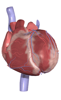
21. Mention the great vessels of the heart with figure.06
22. Describe the short junctional tissue of the heart. 06
23. Describe the heart valve with their position. 07
24. Describe in brief the valves of heart. 08
Diaphragm
25. Write down the main parts and major openings of the diaphragm. 06
Write down the major opening of the diaphragm with their vertebral level. 08
26. Mention the important structures passing through the major opening. 06
Mention the structure passes through them. 08
27. Mention the differences between large and small intestine. 07, 08
28. Write the parts of small intestine with length. 09, 08
Mention the layers of small intestine serially. 08
29. Write down the parts of large intestine with figure. 06
30. Give the branches of abdominal aorta. 06
Female genital organ
31. Write the structure and blood supply of the uterus? 07
Mention the supports and blood supply of uterus. 08
32. State the different parts of uterus with diagram. 09
33. Give the histology of ovary with figure. 09
34. State the function of a fallopian tube. 09
35. Write down the female genital organ with figure. 06
Male genital organ
36. Mention parts of male genital organ with figure. 06
37. Mention the parts of male genital system with figure. 08
38. Write down the layers of scrotum. 08
39. What is prostate? Give its function. 07
40. Write the layers of eyeball. 09
41. Describe visual pathway in brief. 09
42. Write the layers of cornea. 09
43. Give the supply of the tongue. 07
Write down the nerve supply of the tongue. 08
44. Give the intrinsic muscles of the larynx. Write down the structures of its mucosa with nerve supply. 07
45. Describe the middle ear cavity. 07
46. How is the mediastinum divided? Give the boundary and content of the middle midistinum 07
47. Write down in brief about pleura. 08
48. Define the roots of the lung. What are the structures pass through it. 07
49. What is bronchial tree? Give the branches. Give the relation of the mediastinal surface of the right and left lung
50. Mention the parts of alimentary canal. 07
51. Describe the branches of thoracic aorta with diagram. 08
Nephron
52. With picture describe briefly about nephron. 07
Draw and level a nephron. 06
What is nephrone, mention the parts with figure. 08
Nervous system
53. Give the different parts of neuron with diagram. 09
54. Classify nervious system. 09,06
55. What are the organs of special sense? 09, 07
Give their function. 07
56. Define neuron with figure. 06
57. Give the difference between sympathetic and parasympathetic nerves system. 08
58. Mention the cranial nerves with origin type and nature. 08
59. Mention the region of abdomen. Give the structure present in right hypochondriac. 08
60. Describe blood supply of vermiform appendix 07
Short note:
· Meninges 09
· Pleura 09, 06
· Thoracic duct 09, 07
· Islets of langerhans 09
· Porta hepatis 09
· macburney’s point 07
· CSF 07
· fallopian tube 07, 06
· pericardium o8, 07, 06
· cranial nerves 06
· Trans pyloric plane 08. 06
· Neuron 08
· vermiform appendix 08
· spinal cord 08
Dr.jakir hossain
BHMS (dhaka university)
Question of Anatomy 1st paper (2008-2006)
Question of 1st professional B.H.M.S. Exam of Dhaka University
Question of Anatomy 1st paper (2008-2006)
1) Define cell. Draw and level a typical human cell. 09, 07
b. Give the parts of human cell with figure. 06
2) Describe cell membrane and mitochondria. 07
3) Give the function of mitochondria and endoplasmic reticulum. 06
4) What is nucleus? What is chromosome? 08
5) What are cell organells? 08
6) Give short note on endoplasmic reticulum. 08
7) What is chromatin? Give its function. 06
8) What are the types of cell division? Describe prophase 1 of first mitotic cell division. 06
9) What do you mean by horizontal, median, sagital and frontal plane? 06
10) Describe chromosome in brief. 07
11) Give the difference between mitosis and meiosis. 07
12) Describe different stages of mitosis with diagram. 09, 08
13) Define cell membrane .write structures of cell membrane with diagram. 09
14) What is gland classify different types of gland with diagram. 08
15) What are the germ layers? Mention derivatives of Ectoderm. 08, 07, 06
16) a. Define synovial joint. Write down the characteristic of synovial joint. 09
b. Draw and level typical synovial joint. 07, 06
17) Classify bone with example. 09
18) Mention blood supply of long bone. 09, 07
19) Classify joint (06) with example. 09
20) Classify synovial joint with example. 07
21) Draw and level intervertibral joint and write its different functions. 07
22) Write down the bones of lower limb. Draw and level a femur. 06
23) Describe a typical thoracic vertebra with figure. 06
24) Write down the peculiarities of clavicle. 06
25) Give the composition and function of bone. 09, 07
26) Give the parts of growing bone with figure. 07, 06
27) Write in short about intervertibral disc. 09
28) Name the quadriceps and hamstrings muscles with nerve supply.07
29) What are the hamstrings muscles? Give their origin, insertions, nerve supply and actions. 08
30) Write the origin insertion and action of following muscles: (any three) a. biceps brachii 09, 07, 06 b. deltoid 09, 07 c. pectoralis major 09, 07d. sartorius 09, 07, 06 e. rectus femoris 07 f. soleus 07 g. trapezius 06
31) Write the origin, insertions, nerve supply and action of rectus abdominis muscles. 09
32) Name the muscles of mastication (06) with their function. 09
33) Give the difference between artery and vein. 08
34) Give the muscles of anterior abdominal wall. 09 and describe rectus abdominis in short. 07
35) What is hip joint formed. What type of joint it is? Give its functions. 08
36) Define and classify tissue. 09, 07
37) Define tissue. Draw and level different type of connective tissue. 08 Write down the types of connective tissue and connective tissue cells. 07
38) Classify connective tissue. 08
39) Give the characteristic of connective tissue.
40) Classify epithelial tissue. Write it function. 09, 07
41) Differentiate epithelial tissue and connective tissue. 09
42) Name the location where simple columnar epithelium is found. 08
43) Give the characteristic of epithelium. 06
44) Classify muscular tissue with example. 07
45) Differentiate among the muscular tissue 07
46) Define rectus sheath? Give the boundary and contents. 08
47) Mention the layers of skin with figures. 08
48) Define ovulation and fertilization .07
49) Write short notes:
Dr.jakir hossain
BHMS (dhaka university)
Subscribe to:
Posts (Atom)