The vertebrate heart is principally composed of
cardiac muscle and
connective tissue. Cardiac muscle is an involuntary striated muscle tissue found only in this organ and responsible for the ability of the heart to pump blood. The average
human heart, beating at 72 beats per minute, will beat approximately 2.5 billion times during an average 66 year lifespan. It weighs approximately 250 to 300 grams (9 to 11 oz) in females and 300 to 350 grams (11 to 12 oz) in males.
[1]Structure
The structure of the heart varies among the different branches of the
animal kingdom. (See
Circulatory system.)
Cephalopods have two "gill hearts" and one "systemic heart". In vertebrates, the heart lies in the anterior part of the body cavity, dorsal to the gut. It is always surrounded by a
pericardium, which is usually a distinct structure, but may be continuous with the
peritoneum in jawless and cartilaginous fish.
Hagfishes, uniquely among vertebrates, also possess a second heart-like structure in the tail.
[8]
In humans
Main article:
Human heart 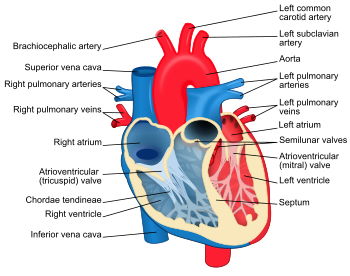
Structure diagram of the human heart from an anterior view. Blue components indicate de-oxygenated blood pathways and red components indicate oxygenated pathways.
The human heart has a mass of between 250 and 350 grams and is about the size of a fist.
[9] It is located anterior to the vertebral column and posterior to the sternum.
It is enclosed in a double-walled sac called the
pericardium. The superficial part of this sac is called the fibrous pericardium. This sac protects the heart, anchors its surrounding structures, and prevents overfilling of the heart with blood.
The outer wall of the human heart is composed of three layers. The outer layer is called the
epicardium, or visceral pericardium since it is also the inner wall of the pericardium. The middle layer is called the
myocardium and is composed of cardiac muscle which contracts. The inner layer is called the
endocardium and is in contact with the blood that the heart pumps. Also, it merges with the inner lining (
endothelium) of blood vessels and covers heart valves.
[10]
The human heart has four chambers, two superior atria and two inferior ventricles. The atria are the receiving chambers and the ventricles are the discharging chambers. The pathway of blood through the human heart consists of a pulmonary circuit
[11] and a systemic circuit. Deoxygenated blood flows through the heart in one direction, entering through the
superior vena cava into the
right atrium and is pumped through the
tricuspid valve into the
right ventricle before being pumped out through the
pulmonary valve to the
pulmonary arteries into the
lungs. It returns from the
lungs through the
pulmonary veins to the
left atrium where it is pumped through the
mitral valve into the
left ventricle before leaving through the
aortic valve to the
aorta.
[12][13]
The fully divided heart
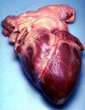
Human heart removed from a 64-year-old man

Surface anatomy of the human heart. The heart is demarcated by:
-A point 9 cm to the left of the
midsternal line (apex of the heart)
-The seventh right
sternocostal articulation
-The upper border of the third right costal cartilage 1 cm from the right
sternal line
-The lower border of the second left costal cartilage 2.5 cm from the left lateral sternal line.
[14] Archosaurs (
crocodilians and
birds) and
mammals show complete separation of the heart into two pumps for a total of four
heart chambers; it is thought that the four-chambered heart of archosaurs evolved independently from that of mammals. In crocodilians, there is a small opening, the
foramen of Panizza, at the base of the arterial trunks and there is some degree of mixing between the blood in each side of the heart; thus, only in birds and mammals are the two streams of blood – those to the pulmonary and systemic circulations – kept entirely separate by a physical barrier.
[8]
In the human body, the heart is usually situated in the middle of the
thorax with the largest part of the heart slightly offset to the left, although sometimes it is on the right (see
dextrocardia), underneath the
sternum. The heart is usually felt to be on the left side because the
left heart (left ventricle) is stronger (it pumps to all body parts). The left
lung is smaller than the right lung because the heart occupies more of the left
hemithorax. The heart is fed by the
coronary circulation and is enclosed by a sac known as the
pericardium; it is also surrounded by the
lungs. The pericardium comprises two parts: the fibrous pericardium, made of
dense fibrous connective tissue, and a double membrane structure (parietal and visceral pericardium) containing a
serous fluid to reduce friction during heart contractions. The heart is located in the
mediastinum, which is the central sub-division of the thoracic cavity. The mediastinum also contains other structures, such as the esophagus and trachea, and is flanked on either side by the right and left pulmonary cavities; these cavities house the
lungs.
[15]
The
apex is the blunt point situated in an inferior (pointing down and left) direction. A
stethoscope can be placed directly over the apex so that the beats can be counted. It is located posterior to the 5th intercostal space just medial of the left mid-clavicular line. In normal adults, the mass of the heart is 250–350 grams (9–12 oz), or about twice the size of a clenched fist (it is about the size of a clenched fist in children), but an extremely diseased heart can be up to 1000 g (2 lb) in mass due to
hypertrophy. It consists of four chambers, the two upper atria and the two lower ventricles.
Functioning
In mammals, the function of the right side of the heart (see
right heart) is to collect de-oxygenated blood, in the
right atrium, from the body (via superior and inferior vena cavae) and pump it, through the tricuspid valve, via the
right ventricle, into the lungs (
pulmonary circulation) so that carbon dioxide can be dropped off and oxygen picked up (
gas exchange). This happens through the passive process of
diffusion. The left side (see
left heart) collects oxygenated blood from the
lungs into the
left atrium. From the left atrium the blood moves to the
left ventricle, through the bicuspid valve (mitral valve), which pumps it out to the body (via the aorta). On both sides, the lower ventricles are thicker and stronger than the upper atria. The muscle wall surrounding the left ventricle is thicker than the wall surrounding the right ventricle due to the higher force needed to pump the blood through the
systemic circulation.
Starting in the right atrium, the blood flows through the
tricuspid valve to the right ventricle. Here, it is pumped out the pulmonary semilunar valve and travels through the pulmonary
artery to the lungs. From there, oxygenated blood flows back through the pulmonary
vein to the left atrium. It then travels through the
mitral valve to the left ventricle, from where it is pumped through the aortic semilunar valve to the
aorta. The aorta forks and the blood is divided between major arteries which supply the upper and lower body. The blood travels in the arteries to the smaller arterioles and then, finally, to the tiny capillaries which feed each cell. The (relatively) deoxygenated blood then travels to the venules, which coalesce into veins, then to the inferior and superior venae cavae and finally back to the right atrium where the process began.
The heart is effectively a
syncytium, a meshwork of cardiac muscle cells interconnected by contiguous cytoplasmic bridges. This relates to electrical stimulation of one cell spreading to neighboring cells.
Some cardiac cells are self-excitable, contracting without any signal from the nervous system, even if removed from the heart and placed in culture. Each of these cells have their own intrinsic contraction rhythm. A region of the human heart called the
sinoatrial (SA) node, or pacemaker, sets the rate and timing at which all cardiac muscle cells contract. The SA node generates electrical impulses, much like those produced by nerve cells. Because cardiac muscle cells are electrically coupled by inter-calated disks between adjacent cells, impulses from the SA node spread rapidly through the walls of the artria, causing both artria to contract in unison. The impulses also pass to another region of specialized cardiac muscle tissue, a relay point called the
atrioventricular node, located in the wall between the right atrium and the right ventricle. Here, the impulses are delayed for about 0.1s before spreading to the walls of the ventricle. The delay ensures that the artria empty completely before the ventricles contract. Specialized muscle fibers called
Purkinje fibers then conduct the signals to the apex of the heart along and throughout the ventricular walls. The Purkinje fibres form conducting pathways called bundle branches. This entire cycle, a single heart beat, lasts about 0.8 seconds. The impulses generated during the heart cycle produce electrical currents, which are conducted through body fluids to the skin, where they can be detected by electrodes and recorded as an
electrocardiogram (ECG or EKG).
[16] The events related to the flow or
blood pressure that occurs from the beginning of one
heartbeat to the beginning of the next can be referred to a
cardiac cycle.
[17]
The SA node is found in all
amniotes but not in more primitive vertebrates. In these animals, the muscles of the heart are relatively continuous and the sinus venosus coordinates the beat which passes in a wave through the remaining chambers. Indeed, since the sinus venosus is incorporated into the right atrium in amniotes, it is likely
homologous with the SA node. In teleosts, with their vestigial sinus venosus, the main centre of coordination is, instead, in the atrium. The rate of heartbeat varies enormously between different species, ranging from around 20 beats per minute in
codfish to around 600 in
hummingbirds.
[8]
Cardiac arrest is the sudden cessation of normal heart rhythm which can include a number of pathologies such as
tachycardia, an extremely rapid heart beat which prevents the heart from effectively pumping blood, which is an irregular and ineffective heart rhythm, and
asystole, which is the cessation of heart rhythm entirely.
Cardiac tamponade is a condition in which the fibrous sac surrounding the heart fills with excess fluid or blood, suppressing the heart's ability to beat properly. Tamponade is treated by
pericardiocentesis, the gentle insertion of the needle of a syringe into the
pericardial sac (avoiding the heart itself) on an angle, usually from just below the
sternum, and gently withdrawing the tamponading fluids.